
Specular Microscope
※For availability in your country, please contact your distributor.

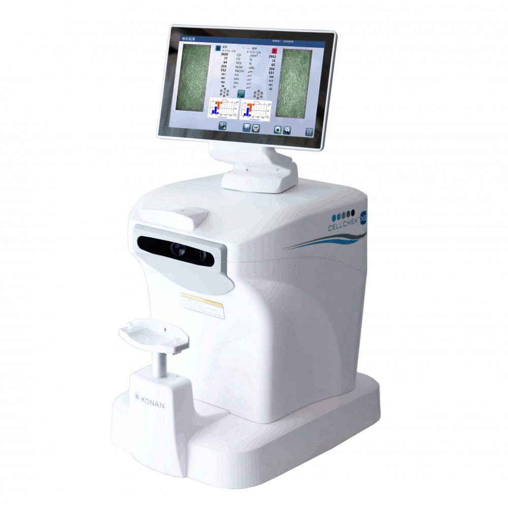
Product name: Konan Specular Microscope XX
Equipped with “Auto Center Method” and “Auto F Center Method” as well as “Auto Trace Method” as standard!
| CellChek 20-1 ・Simple to use fully automated OD/OS endothelium capturing,analysis, printing and exporting, with a touch of screen. ・Enhanced Image Capturing Capability ・Automated Capturing Retry Feature ・Flexible Monitor Direction |
 |
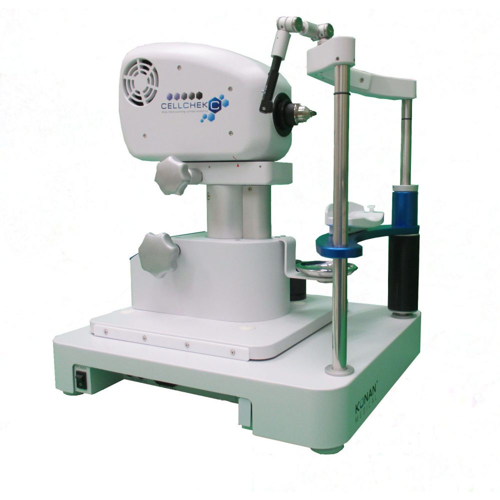
Product name: Konan Specular Microscope XVI
High-grade model providing wide-field visualization of the cornea; including from limbus to limbus, endothelium to epithelium
| CellChek C ・High-resolution / Wide-field ・Full corneal layers visualization ・Desired portion of cornea can be observed ・Analysis software integrated: Auto-trace / Center Method / Flex Center Method ▶▶More Features→ ※Built-to-order model. Please contact us for delivery time. |
 |
![]()
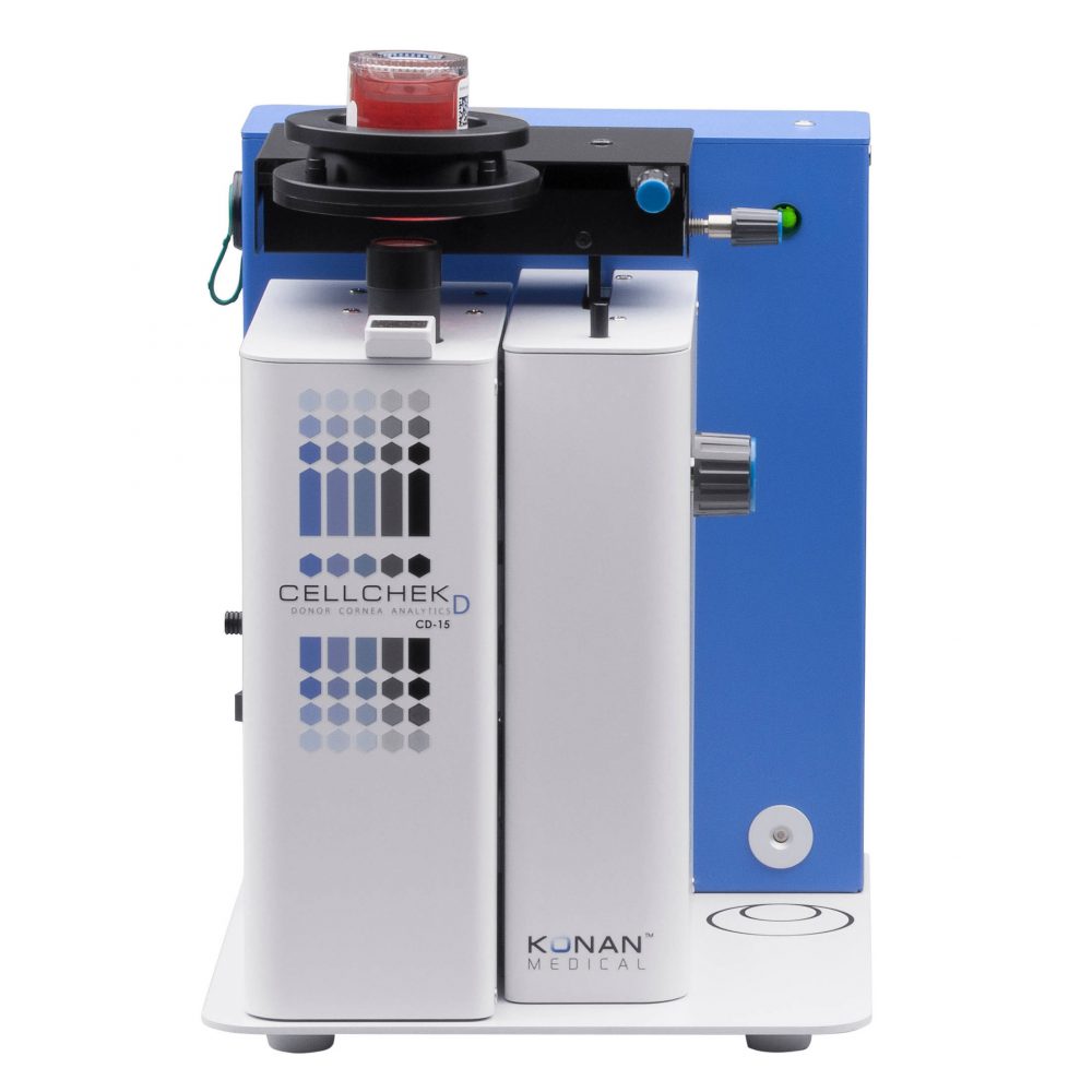
Introducing the new generation of the gold standard quality imaging and assessment of transplantable corneal material.
| CellChek D ・Ultra-wide viewing areas: 1000 x 750μm ・Dual Camera System ・Built-in Pachymeter, real-time media temperature sensor ・Integrated database ・Adaptable to most of the commercially available chambers ※This is upgradable to full features on Konan’s CellChekD Plus. |
 |
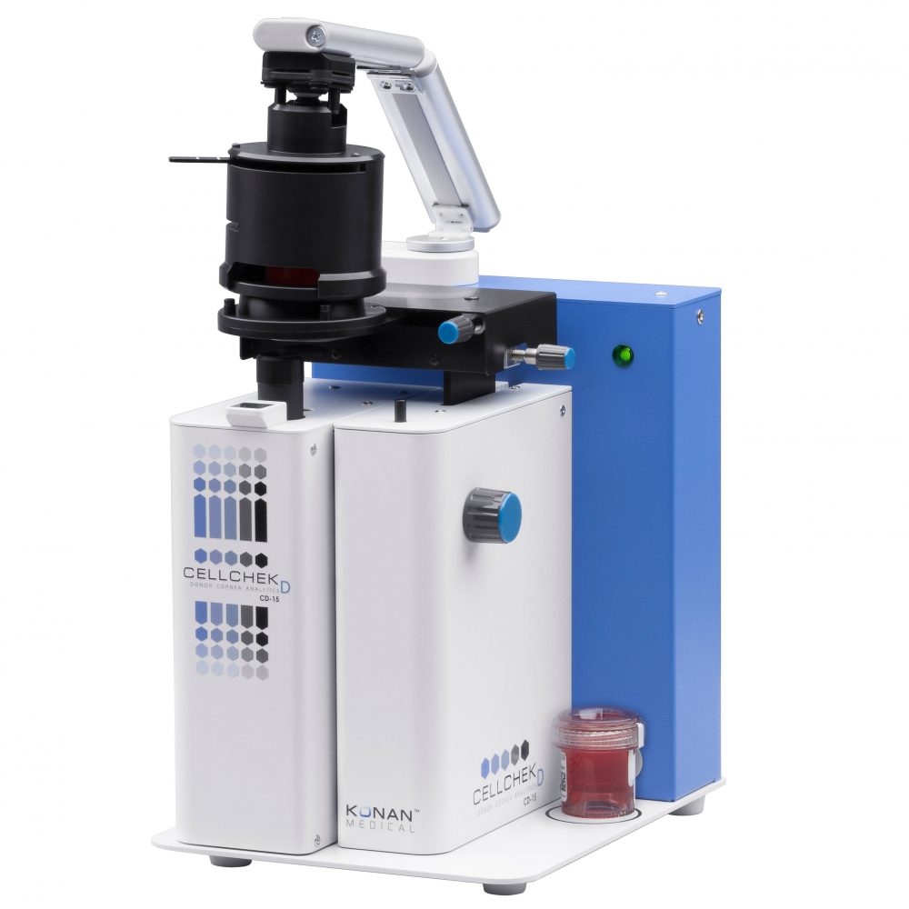
The first multi-imaging system for donor corneal analysis which provides an amazing view into the cornea that simply has never been seen before.
| CellChek D PLUS ・New “Donor Enhance” Imaging system ・Full Graft Imaging & digital measurement tools |
 |