Wide-field Scanning Specular Microscope
Not cleared for sale in the USA.
CellChek C
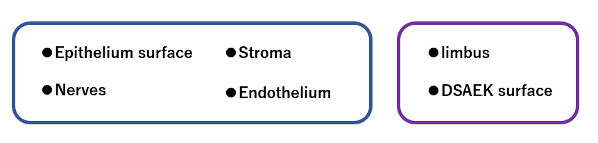
High-grade model providing wide-field visualization of the cornea; including from limbus to limbus, endothelium to epithelium
| ◆ High-resolution / Wide-field ◆ Full corneal layers visualization ◆ Desired portion of cornea can be observed ◆ Analysis software integrated: Auto-trace / Center Method / Flex Center Method | 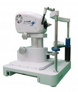 |
※Built-to-order model. Please contact us for delivery time.
| ◆ High-resolution / Wide-field | |||||
| Slit-scanning technology leads to high-res & high-contrast imaging of cornea layers. High quality visualization of corneal endothelium. Extra wide-field: 0.65 mm x 0.48mm. |
|||||
| ◆ Full corneal layers visualization | |||||
|
|||||
| ◆ Desired portion of cornea can be observed | |||||
| Floating mechanism maintains focus distance. Observation location can be moved around from limbus to limbus. Real time movie can be recorded.
|
|||||
| ◆ Analysis software | |||||
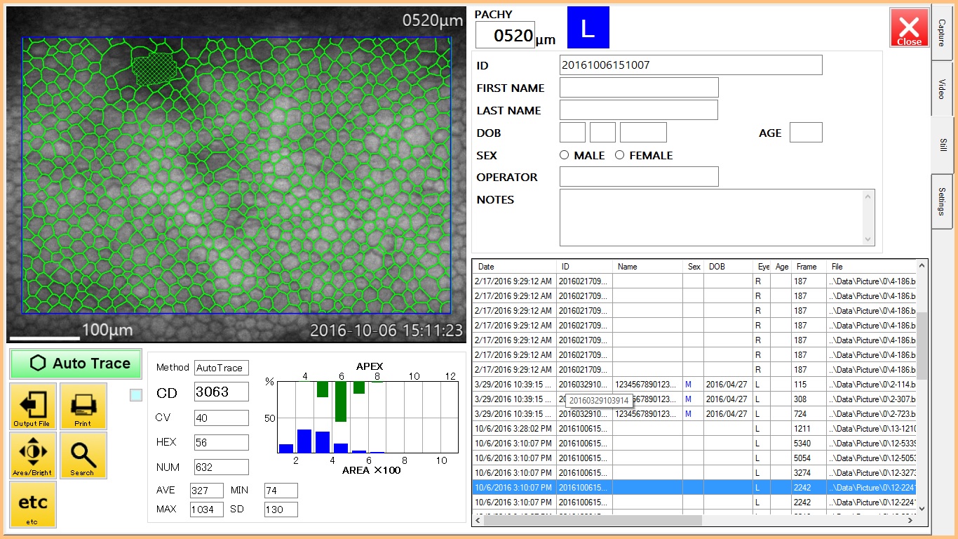
| Exclusive software for corneal endothelium cell analysis with Auto-trace function is integrated. Features a max. 60 min. video imaging system with “stop-action” still images. Videos and still images are saved automatically. Patient database integrated.
|
|||||
| ◆ Specification | |||||
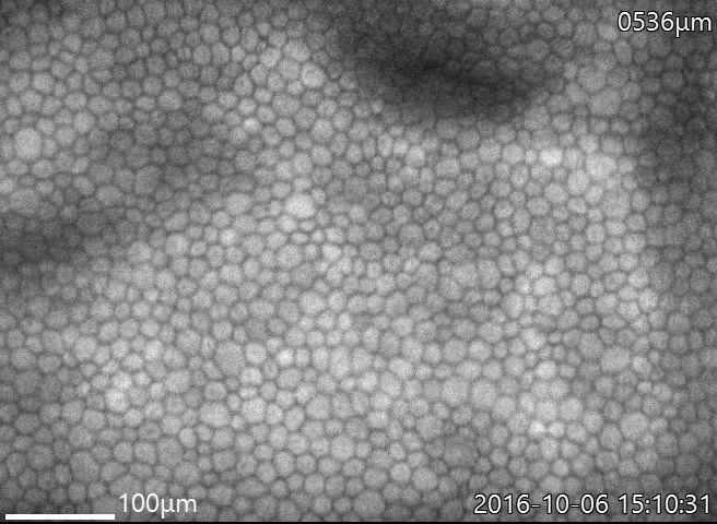 Sample image : Corneal endothelium(central)
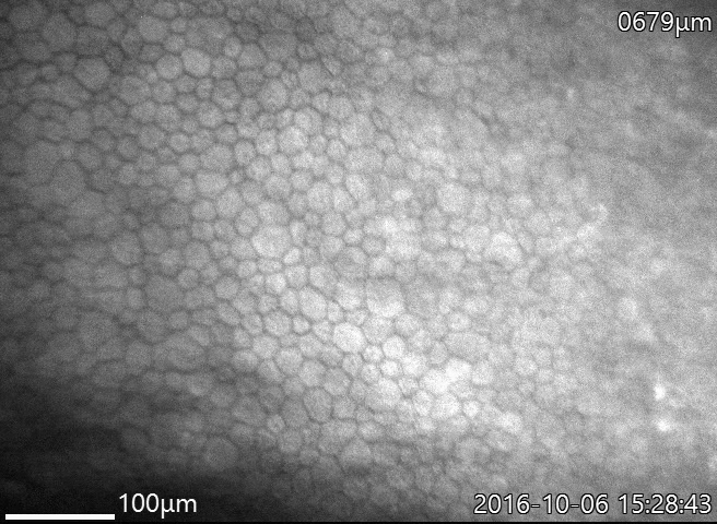 Sample image: Corneal endothelium (near limbus)
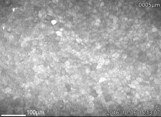 Sample image: Corneal Epithelium surface
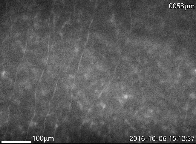 Sample image: Nerves
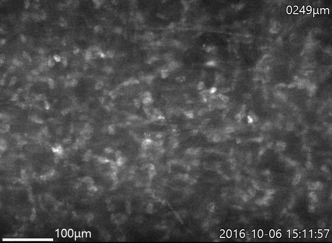 Sample image: Stroma